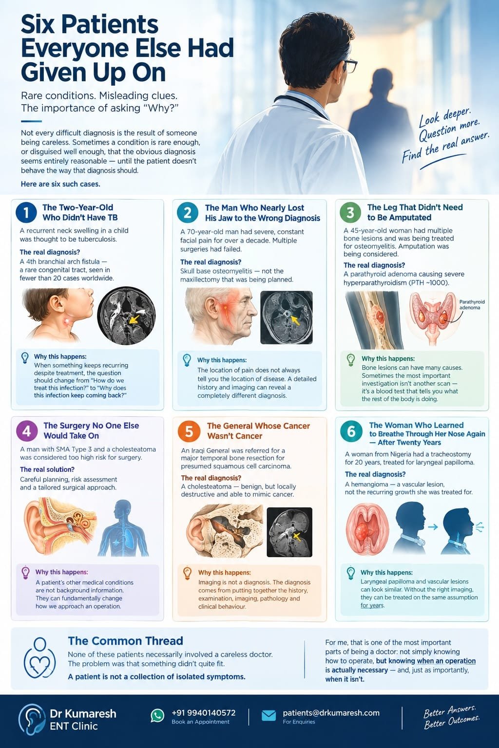
Second Opinion – Six Patients Everyone Else Had Given Up On
Not every difficult diagnosis is the result of someone being careless. Sometimes a condition is rare enough, or disguised well enough, that the obvious diagnosis seems entirely reasonable — until the patient doesn’t behave the way that diagnosis should.
Over the years, I have seen patients who had already undergone procedures, taken prolonged courses of medication, or been advised major surgery — only to discover that the original diagnosis did not fully explain what was happening.
These are six such cases.
What stayed with me was not simply that the diagnoses were unusual. It was what could have happened if we had stopped questioning them.
1. The Two-Year-Old Who Didn’t Have TB
A two-year-old boy had been suffering from a swelling in his neck for nearly six months.
He had already undergone incision and drainage, but the swelling kept coming back. In a child in India with a persistent or recurrent neck abscess, tuberculosis is an entirely understandable diagnosis to consider, and the working diagnosis eventually became a TB neck abscess.
But something about the pattern didn’t fit, I wanted to understand why the problem was recurring. I ordered an MRI.
The scan told a different story.
This wasn’t tuberculosis. It was a 4th branchial arch fistula — a rare congenital tract and including this less than 20 have been reported in the world.
Why this happens
A recurrent neck abscess in a child will quite reasonably be treated as an infection. In India, tuberculosis also has to remain in the differential diagnosis.
But when something keeps recurring despite appropriate treatment, the question should change from:
“How do we treat this infection?”
to:
“Why does this infection keep coming back?”
2. The Man Who Nearly Lost His Jaw to the Wrong Diagnosis
A 70-year-old man came with severe, constant pain on one side of his head and jaw. He described it as 10 out of 10.
It had been more than a decade of this pain. He had already undergone multiple surgeries in search of an answer, but the pain persisted.
The next major procedure being considered was a maxillectomy — removal of part of the jaw and midface — because of the severity and persistence of his pain.
Fortunately, a doctor who had encountered a similar case referred him to me before that operation.
When I reviewed his history and investigations, another possibility became apparent: skull base osteomyelitis.
Once the diagnosis was established and appropriate treatment started, the response was remarkable.
Within four months, he was pain-free.
Why this happens
The location of pain does not always tell you the location of disease.
That is why a detailed history matters so much. The character, duration, progression and distribution of pain — combined with examination and imaging — can sometimes reveal a completely different diagnosis from the one suggested by the location of the symptom alone.
3. The Leg That Didn’t Need to Be Amputated
A 45-year-old woman travelled from another state with severe pain in her leg.
She had multiple bone lesions and had been treated for osteomyelitis. Unfortunately, the lesions were not responding as expected, and amputation was being considered.
A colleague was not convinced that the entire picture fitted osteomyelitis and referred her for another opinion.
That second look changed everything.
The actual problem was a large parathyroid adenoma causing severe hyperparathyroidism.
Her parathyroid hormone level was extraordinarily high — around 1000.
The excessive parathyroid hormone was profoundly affecting her bones, producing lesions and bone pain that had been interpreted as infection.
This is a recognised but now relatively uncommon manifestation of severe hyperparathyroidism: osteitis fibrosa cystica, sometimes associated with so-called brown tumours.
The adenoma was removed.
Her PTH subsequently returned to normal.
And the leg did not need to be amputated.
Why this happens
Bone lesions can have many causes.
If the imaging is considered in isolation, infection or malignancy may seem like the obvious possibilities. But the patient is not an X-ray, CT or MRI.
Sometimes the most important investigation for a bone lesion isn’t another scan. It is a blood test that tells you what the rest of the body is doing.
4. The Surgery No One Else Would Take On
A man from another state had a particularly challenging combination: Spinal Muscular Atrophy (SMA) Type 3 and a cholesteatoma.
The cholesteatoma was destructive and required surgery.
But the problem was not simply the ear.
SMA meant significant muscle weakness and increased anaesthetic and respiratory concerns. A prolonged operation and prolonged anaesthesia could potentially carry substantial risks, particularly in the postoperative period.
Understandably, several surgeons were reluctant to take on the case.
A colleague eventually told him that there was one surgeon he should consult, and he travelled to see me.
The important point was that this was not a situation where the risks could simply be ignored.
They had to be understood, assessed and planned around.
After the necessary preoperative evaluation, the surgical strategy was designed with the patient’s underlying condition in mind. The objective was not merely to perform the operation, but to do it efficiently while minimising unnecessary operative and anaesthetic exposure.
The surgery was completed safely.
The patient recovered well and is doing well today.
Why this matters
A patient’s other medical conditions are not background information.
They can fundamentally change how we approach an operation.
A procedure that is routine in one patient may require completely different planning in another.
The answer to increased risk isn’t always to avoid surgery.
Sometimes it is to understand the risk properly and change the way the surgery is performed.
5. The General Whose Cancer Wasn’t Cancer
An Iraqi General was referred to me for a temporal bone resection.
The presumed diagnosis was squamous cell carcinoma of the temporal bone — a serious diagnosis for which radical surgery may be necessary.
But when I reviewed his history and imaging, something didn’t quite fit.
I suspected that we needed to revisit the diagnosis before proceeding with such a major operation.
The workup was repeated.
It wasn’t cancer.
It was a cholesteatoma.
Cholesteatoma is benign in the sense that it is not a cancer, but it can be locally destructive and erode bone. In certain situations, its appearance can mimic a malignant temporal bone tumour.
The distinction was critical.
The patient avoided a radical cancer operation that he did not need.
He kept his face.
And he is doing well today.
Why this happens
Imaging can be incredibly powerful.
But imaging is not a diagnosis.
The diagnosis comes from putting together the history, examination, imaging, pathology and clinical behaviour of the disease.
When the proposed treatment is irreversible or carries major consequences, there is enormous value in asking one simple question:
“Are we absolutely certain this is what we are treating?”
6. The Woman Who Learned to Breathe Through Her Nose Again — After Twenty Years
A woman from Nigeria had been living with a tracheostomy for more than twenty years.
She had originally been diagnosed with laryngeal papilloma, and had undergone repeated procedures over the following two decades to manage it. The tracheostomy tube — breathing through an opening in her neck rather than her nose and mouth — had become simply how she lived.
Eventually, her condition reached a point where it could no longer be managed the way it had been. She was referred for another opinion.
A contrast MRI told a very different story.
This was not papilloma. It was a hemangioma — a vascular lesion, not the recurring growth she had been treated for, for twenty years.
Once correctly identified, it was treated successfully.
The tracheostomy tube — the one she had lived with for more than two decades — was removed.
After twenty years, she learned to breathe through her natural airway again. Her nose.
Her smile, and her gratitude, were the real measure of what that meant.
Why this happens
Laryngeal papilloma and vascular lesions like hemangioma can sit in similar territory and, without the right imaging, get treated on the same assumption for years — especially once a patient has settled into repeated procedures for a presumed diagnosis, and the question of whether it’s the right diagnosis stops being asked.
Twenty years is a long time to breathe through a tube for a diagnosis that was never quite right. It is also a reminder that “long-standing” and “confirmed” are not the same thing.
The Common Thread
Looking back at these six patients, there is one thing that connects them.
None of them necessarily involved a careless doctor.
In fact, many of the initial diagnoses were entirely understandable.
The problem was that something didn’t quite fit.
And that is precisely why I spend considerable time eliciting the history and looking at the patient as a whole, rather than treating only the symptom that brought them to me.
A patient is not a collection of isolated symptoms.
For me, that is one of the most important parts of being a doctor: not simply knowing how to operate, but knowing when an operation is actually necessary — and, just as importantly, when it isn’t.