Pulsatile Tinnitus: Why That Whooshing Sound in Your Ear Might Not Be Dangerous
If you’ve ever noticed a rhythmic whooshing, swishing or thumping sound in your ear — one that seems to beat in time with your heart — you’ve likely already done what most people do next: searched it online, found a list of frightening possibilities, and panicked.
Before you spiral through worst-case scenarios, let’s go through this properly. What pulsatile tinnitus actually is, why carotid canal dehiscence is one of the most common and most reassuring explanations, what other causes need to be ruled out, and what tests and treatment actually look like.
What makes pulsatile tinnitus different from ordinary tinnitus?
Most tinnitus is a constant ringing, hissing or buzzing sound that has nothing to do with your heartbeat.
Pulsatile tinnitus is different. It’s rhythmic, and it typically beats in sync with your pulse. That single feature — the synchronicity with your heartbeat — is what tells us this is very likely coming from blood flow somewhere near the ear, rather than from the hearing nerve itself.
That distinction matters enormously, because it points us toward a specific set of causes rather than a vague, hard-to-pin-down symptom.
Carotid Canal Dehiscence — the reassuring explanation you need to hear about
This is one of the most common findings we identify when we investigate pulsatile tinnitus, and it deserves to be explained clearly because it is frequently misunderstood as something dangerous when it usually isn’t.
The carotid artery — the major blood vessel supplying your brain — passes very close to the middle ear as it travels up through the skull, inside a bony channel called the carotid canal. Normally, a layer of bone separates this artery from the middle ear space.
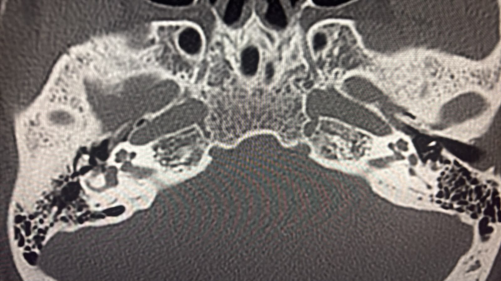
In carotid canal dehiscence, that bony covering is thin or, in some people, focally absent — a variation in anatomy that some people are simply born with. Because the artery sits that much closer to the middle ear, the normal turbulence of blood flow within it becomes audible as a pulsing sound.
Here is the reassurance: carotid canal dehiscence is, in the overwhelming majority of cases, a benign anatomical variant. It is not a sign of a growing problem, it is not typically progressive, and it does not usually indicate any danger to the artery itself. Many people live with it as an incidental finding, identified on imaging done specifically to investigate the tinnitus, with no other consequence.
This is not the same as an aneurysm, and it is not the same as a tumor. It is, most often, simply an anatomical variation that explains why you’re hearing what you’re hearing — and once identified and confirmed benign, the appropriate next step is often reassurance and monitoring rather than intervention.
Other causes of pulsatile tinnitus
Carotid canal dehiscence is common, but it isn’t the only explanation, and part of a proper evaluation is making sure it isn’t something else. Other recognized causes include:
- Atherosclerosis or carotid artery narrowing — turbulent flow through a narrowed vessel can produce a similar sound, particularly in older patients or those with vascular risk factors
- Venous causes — an abnormally high jugular bulb, or a small outpouching in the wall of the sigmoid sinus (a sigmoid sinus diverticulum or dehiscence), can both produce pulsatile sound from the venous side rather than the arterial side
- Idiopathic intracranial hypertension — raised pressure around the brain, more common in young women, can present with pulsatile tinnitus, often alongside headache or visual symptoms
- Dural arteriovenous fistula — an abnormal connection between arteries and veins near the brain; this is one of the more serious causes and is specifically why proper imaging matters rather than assuming every case is benign
- Glomus tumors (paragangliomas) — vascular tumors that can arise in the middle ear or at the skull base; less common, but an important reason to have persistent pulsatile tinnitus formally evaluated
- High-flow states — anemia or an overactive thyroid can increase blood flow enough to make normal vessels audible, even without any structural abnormality
The point of this list isn’t to alarm you — it’s to explain why a proper evaluation is worthwhile, precisely so that a benign cause like carotid canal dehiscence can be confirmed, rather than assumed.
What tests are actually used?
A structured evaluation typically includes:
- A detailed clinical examination, including otoscopy and listening over the neck, mastoid and orbit with a stethoscope — this alone can sometimes suggest whether the sound is arterial or venous in origin
- A hearing test (audiogram), to check whether there’s any associated hearing change
- High-resolution CT of the temporal bone, which is particularly useful for identifying carotid canal dehiscence, as well as other bony abnormalities near the middle ear
- MRI/MRA or CT angiography, to assess the blood vessels directly and rule out more significant vascular causes such as a dural fistula or aneurysm
- Blood tests, such as a hemoglobin level or thyroid function, when a high-flow state is suspected as a contributing factor
Not every patient needs every test — the specific combination depends on what the examination suggests.
How is it managed?
Management depends entirely on what’s found — which is exactly why the evaluation matters.
- If carotid canal dehiscence is confirmed and no other abnormality is identified, management is often simply reassurance and monitoring. No surgery, no intervention — just an explanation of what’s causing the sound and confirmation that it isn’t dangerous.
- If an underlying contributor like anemia or thyroid dysfunction is found, treating that condition often improves or resolves the symptom.
- If idiopathic intracranial hypertension is the cause, management is directed at reducing intracranial pressure, usually in coordination with neurology.
- If a dural arteriovenous fistula or similar vascular abnormality is identified, referral for specialist vascular or neurosurgical management is appropriate.
- If a glomus tumor is found, management is planned individually, often surgically, depending on its size and location.
When should you see a specialist?
You should have pulsatile tinnitus formally evaluated — rather than simply living with the worry — particularly if:
- The sound is persistent rather than brief or occasional
- It’s associated with hearing loss, headache, visual changes, or dizziness
- It started suddenly or has changed in character
- You have risk factors for vascular disease
The bottom line
Hearing a whooshing or pulsing sound in your ear is unsettling, and it’s reasonable to want an answer rather than to just live with it. But most causes of pulsatile tinnitus, once properly evaluated, turn out to be benign — and carotid canal dehiscence in particular is one of the most common and most reassuring explanations we find.
The goal of evaluation isn’t to alarm you. It’s to confirm what’s actually happening, rule out the small number of causes that do need treatment, and — in most cases — give you the reassurance that comes from actually knowing rather than guessing.