Carotid Body Tumour: A Rare Tumour That Demands Specialised Surgical Expertise
A Carotid Body Tumour (CBT) is a rare tumour arising from the carotid body, a small structure located at the division of the common carotid artery into the internal and external carotid arteries. These tumours are also known as carotid paragangliomas, since the carotid body itself is a paraganglion — a cluster of chemoreceptor cells that monitors oxygen and carbon dioxide levels in the blood.
Although they are uncommon, I have personally had the opportunity to manage three patients with Carotid Body Tumours, all of whom came to me specifically for surgical treatment.
These cases are a reminder that, when a tumour develops in an anatomically complex location, the outcome depends not only on identifying the disease but also on having the appropriate surgical training, experience and preparedness to deal with the challenges that may arise during surgery.
Why is a Carotid Body Tumour challenging?
The carotid body lies at an extremely important anatomical location, immediately adjacent to the major blood vessels supplying the brain and surrounded by important nerves.
As the tumour grows, it can gradually surround or displace the carotid arteries and may also become closely related to the cranial nerves in the neck.
This makes surgery technically demanding.
The surgeon has to balance several objectives:
- Complete and safe tumour removal
- Preservation of blood flow to the brain
- Protection of the cranial nerves
- Control of major blood vessels if necessary
- Minimising the risk of neurological complications
The difficulty can increase substantially when the tumour has become intimately associated with the carotid artery.
What causes a Carotid Body Tumour?
The carotid body is a chemoreceptor organ, and most known risk factors for a tumour developing in it relate directly to how that organ functions:
- Chronic hypoxia — long-term low oxygen exposure, classically seen in people living at high altitude, is associated with enlargement of the carotid body and eventual tumour formation.
- Hereditary paraganglioma syndromes — a meaningful proportion of cases, particularly bilateral ones, are linked to inherited mutations in the succinate dehydrogenase (SDH) genes (SDHB, SDHC, SDHD). A family history of paraganglioma or pheochromocytoma should raise suspicion.
- Sporadic occurrence — the majority of patients have no identifiable hereditary cause; a solitary carotid body tumour simply arises on its own.
This is why family history is always part of my assessment, even when a case looks straightforward on imaging.
One of my cases required carotid artery reconstruction
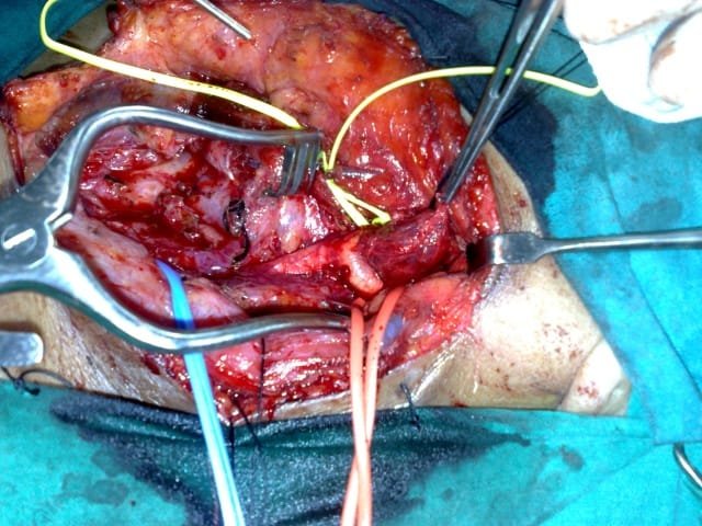
In one of the three patients I operated on, the tumour was so closely associated with the carotid artery that it was necessary to divide the carotid artery during tumour removal and subsequently perform an anastomosis of the divided ends to restore continuity of the artery.
This is a highly specialised surgical situation.
It requires careful preoperative assessment, detailed knowledge of vascular and Head & Neck anatomy, meticulous dissection and the ability to perform vascular reconstruction when required.
Such cases also highlight an important principle in complex surgery: the surgeon must be prepared for what the anatomy demands, rather than simply following a predetermined surgical plan.
A Word of Caution: What Not To Do
A case that stays with me. I once had a patient referred to me after a needle biopsy (FNAC) had been performed elsewhere for what was assumed to be a routine neck swelling. What followed was a near-fatal bleed. By the time he reached me, he was weak, and his neck had turned visibly blue from the extent of the internal bleeding.
A Carotid Body Tumour is not a routine neck lump. It is one of the most vascular masses that can occur in the neck, sitting directly on the carotid vessels. Needling it blindly, without first knowing what you are dealing with, can trigger bleeding that is very difficult to control outside a controlled surgical setting.
This is why I insist on an ultrasound, at minimum, before anything is put into a neck swelling — and why, when the ultrasound or clinical picture suggests a vascular lesion, I go further with contrast imaging before any needle ever comes near it.
A few principles follow directly from this:
- Do not perform FNAC or needle biopsy on a suspected vascular neck mass without prior imaging to characterise it. This is the single most important precaution.
- Do not assume a slow-growing, painless neck lump is “nothing to worry about.” Carotid Body Tumours are often painless for a long time and can still be significant.
- Do not delay imaging once a pulsatile or unusually firm neck swelling is noticed, even if the patient feels otherwise well.
Why specialised training matters
Complex Head & Neck surgery is not simply about removing a tumour.
It involves understanding the relationship between the tumour and the major vessels, nerves, airway and other critical structures and being prepared to manage unexpected findings during surgery.
My training has included specialised Head & Neck Surgery and Oncology at Roswell Park, one of the leading cancer centres in the world, followed by advanced training in Otology and Neurotology at the University of Cincinnati.
This combination of extensive Head & Neck surgical training and experience with complex anatomy has been particularly valuable in managing uncommon and technically demanding conditions.
When dealing with a rare tumour such as a Carotid Body Tumour, this experience becomes especially important because there may be relatively few opportunities for a surgeon to develop familiarity with these cases.
Early diagnosis and appropriate evaluation are important
A Carotid Body Tumour may present as a slowly growing, usually painless lump in the upper part of the neck. Some patients may have very few symptoms despite the tumour reaching a considerable size.
Imaging plays an important role in diagnosis and surgical planning. Contrast-enhanced CT, MRI and vascular imaging can help determine the size of the tumour and its relationship to the carotid arteries and surrounding structures.
The extent to which the tumour surrounds the carotid vessels is also important when assessing the complexity and risks of surgery.
When should you see a specialist?
Certain features of a neck lump should prompt a specialist evaluation before any procedure is attempted on it:
- A neck lump that is pulsatile, or where you can feel a throb in sync with the heartbeat
- A lump that moves side to side but not up and down on swallowing — a classic sign for masses in this location
- An audible whooshing sound (bruit) over the swelling
- Symptoms such as hoarseness, tongue weakness, or difficulty swallowing, suggesting cranial nerve involvement
- A family history of paraganglioma or pheochromocytoma
- Any neck swelling in the upper neck near the jaw angle that is slowly enlarging, regardless of whether it is painful
If any of these apply, the right first step is imaging and a specialist opinion — not a needle.
Surgery requires planning, precision and experience
The treatment of a Carotid Body Tumour needs to be individualised according to the size of the tumour, its relationship to the carotid vessels, involvement of adjacent nerves, the patient’s overall health and other clinical factors.
For larger or more advanced tumours, surgery may require collaboration between Head & Neck surgeons, vascular surgeons, radiologists, anaesthetists and other specialists.
In selected patients, preoperative planning may also include assessment of cerebral circulation and consideration of vascular reconstruction strategies.
The goal is not merely to remove the tumour.
The goal is to remove it safely while preserving the function of the brain, major blood vessels and cranial nerves whenever possible.
Three patients, three reminders of the importance of expertise
Having treated three patients with Carotid Body Tumours, including one in whom carotid artery division and reconstruction was necessary, I am reminded of why specialised surgical training matters.
Rare and complex diseases require more than familiarity with the diagnosis. They require the ability to understand difficult anatomy, anticipate complications, make decisions under pressure and have the technical skills necessary to manage those situations safely.
When the anatomy is complex, surgical expertise can make the difference.
If you or someone you know has been diagnosed with a Carotid Body Tumour or has a slowly growing lump in the upper neck, an evaluation by a specialist experienced in Head & Neck surgery is important.
This article is intended for general information and does not replace an individual consultation and appropriate imaging evaluation.